Endoplasmic Reticulum
Key Roles
-
Protein Synthesis and Folding:
- The RER is studded with ribosomes on its cytosolic surface, which synthesize proteins, particularly those that are secreted from the cell, embedded in the cell membrane, or sent to an organelle like the lysosome. This includes hormones, enzymes, and structural proteins.
- After synthesis, the RER also plays a role in the initial folding and post-translational modifications of these proteins, such as glycosylation.
-
Quality Control:
- The RER is crucial for ensuring that newly synthesized proteins are correctly folded. Misfolded proteins are targeted for degradation to prevent cellular damage.
-
Lipid Synthesis:
- While the smooth ER is primarily involved in lipid synthesis, the RER also contributes to membrane phospholipid and cholesterol production, which is essential for maintaining cellular structure and function.
Clinical Significance:
-
Diseases Related to Protein Misfolding (e.g., Cystic Fibrosis, Alzheimer's Disease):
- Defective protein folding or a malfunction in the RER's quality control system can lead to the accumulation of misfolded proteins, which can cause diseases. For example, cystic fibrosis is caused by a misfolded CFTR protein (a chloride channel), and Alzheimer's disease is associated with misfolded amyloid-beta proteins.
- In these conditions, the RER's inability to properly fold proteins can lead to cellular stress, triggering pathways like the unfolded protein response (UPR), which, if prolonged or excessive, can result in cell death.
-
Endoplasmic Reticulum Stress and the Unfolded Protein Response (UPR):
- When the RER is overwhelmed with the accumulation of misfolded proteins, it activates a cellular response known as the UPR. While the UPR initially helps the cell cope by increasing protein chaperones and reducing protein synthesis, chronic stress can lead to cell dysfunction and contribute to diseases such as neurodegeneration, diabetes, and cancer.
-
Congenital Disorders of Glycosylation (CDG):
- These are a group of rare genetic disorders caused by defects in the enzymes involved in glycosylation (a process carried out in part by the RER). Glycosylation errors affect protein function and stability, leading to a variety of systemic symptoms, including developmental delays and organ dysfunction.
-
Toxins and Viral Infections:
- Some pathogens, such as viruses, target the RER to hijack the cell’s protein-synthesis machinery for their replication. Additionally, toxins like ricin can inhibit the function of the ribosomes on the RER, disrupting protein synthesis and leading to cell death.
-
Cancer:
- The RER is involved in the production of proteins required for cell growth and survival. In cancer cells, altered protein synthesis and stress responses may lead to an increased demand on the RER, contributing to tumorigenesis and metastasis.
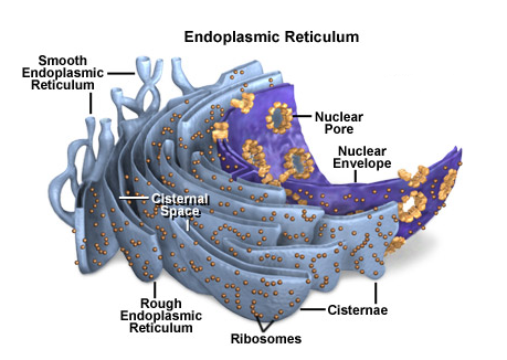
The endoplasmic reticulum is a network of flattened sacs and branching tubules that extends throughout the cytoplasm.
These sacs and tubules are all interconnected by a single continuous membrane so that the organelle has only one large, highly convoluted and complexly arranged lumen (internal space).
It is usually referred to as the endoplasmic reticulum cisternal space.1
The endoplasmic reticulum membrane allows molecules to be selectively transferred between the lumen and the cytoplasm, and since it is connected to the double-layered nuclear envelope, it further provides a pipeline between the nucleus and the cytoplasm.
The endoplasmic reticulum manufactures, processes, and transports a wide variety of biochemical compounds for use inside and outside of the cell. Consequently, many of the proteins found in the cisternal space of the endoplasmic reticulum lumen are there only transiently as they pass on their way to other locations. Other proteins, however, are targeted to constantly remain in the lumen and are known as endoplasmic reticulum resident proteins. These special proteins, which are necessary for the endoplasmic reticulum to carry out its normal functions, contain a specialized retention signal consisting of a specific sequence of amino acids that enables them to be retained by the organelle. An example of an important endoplasmic reticulum resident protein is the chaperone protein known as BiP(formally: the chaperone immunoglobulin-binding protein), which identifies other proteins that have been improperly built or processed and keeps them from being sent to their final destinations.
There are two basic kinds of endoplasmic reticulum morphologies: rough and smooth.
These proteins are then transferred to the Golgi apparatus for further transport either to the cell membrane for secretion or to the lysosomes for degradation. |
|
| ▶ | Location: Found in high concentrations in small intestine and antibody producing plasma cells. |
The surface of rough endoplasmic reticulum is covered with ribosomes, giving it a bumpy appearance when viewed through the microscope. This type of endoplasmic reticulum is involved mainly with the production and processing of proteins that will be exported, or secreted, from the cell. The ribosomes assemble amino acids into protein units, which are transported into the rough endoplasmic reticulum for further processing. These proteins may be either transmembrane proteins, which become embedded in the membrane of the endoplasmic reticulum, or water-soluble proteins, which are able to pass completely through the membrane into the lumen. Those that reach the inside of the endoplasmic reticulum are folded into the correct three-dimensional conformation, as a flattened cardboard box might be opened up and folded into its proper shape in order to become a useful container. Chemicals, such as carbohydrates or sugars, are added, then the endoplasmic reticulum either transports the completed proteins to areas of the cell where they are needed, or they are sent to the Golgi apparatus for further processing and modification.
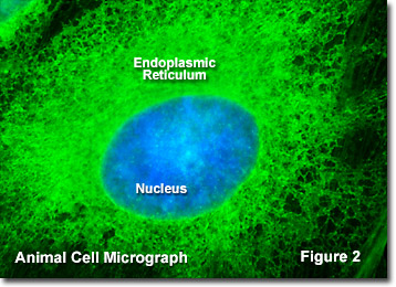
Most proteins exported from the endoplasmic reticulum exit the organelle in vesicles budded from the smooth portion, which has a more even appearance than rough endoplasmic reticulum when viewed through the electron microscope because of the lack of ribosomes. The smooth endoplasmic reticulum in most cells is much less extensive than the rough endoplasmic reticulum and is sometimes alternatively termed transitional. Smooth endoplasmic reticulum is chiefly involved, however, with the production of lipids (fats), building blocks for carbohydrate metabolism, and the detoxification of drugs and poisons. Therefore, in some specialized cells, such as those that are occupied chiefly in lipid and carbohydrate metabolism (brain and muscle) or detoxification (liver), the smooth endoplasmic reticulum is much more extensive and is crucial to cellular function. Smooth endoplasmic reticulum also plays a role in various cellular activities through its storage of calcium and involvement in calcium metabolism. In muscle cells, smooth endoplasmic reticulum releases calcium to trigger muscle contractions. Presented in Figure 2 is a fluorescence digital image taken through the microscope of the endoplasmic reticulum network in a bovine (cow) pulmonary artery endothelial cell grown in culture.
It is the rough endoplasmic reticulum that is directly continuous with the nuclear envelope (as illustrated in Figure 1), which is also studded with ribosomes, and the two organelles are thought to have evolved simultaneously in ancient cells. Due to their physical membranous connection, the lumen of the endoplasmic reticulum and the space between the layers of the nuclear envelope comprise a single compartment. Accordingly, the nucleus has direct access to proteins (many of which are produced by the ribosomes upon its surface) and other materials present in the endoplasmic reticulum lumen, so that transport vesicles are not needed to obtain them. The close association between the endoplasmic reticulum and the nucleus also enables the organelles to share information in a very efficient manner. For instance, if the endoplasmic reticulum begins to undergo functional problems and unfolded proteins accumulate within the organelle, which can be extremely hazardous to the cell, the organelle quickly sends a signal to the nucleus (as well as to the cytoplasm). The nucleus responds by slowing ribosomal translation through a several-step process, thereby giving the endoplasmic reticulum extra time to catch up on its protein folding, thus maintaining cellular health.
++++++
Th endoplasmic reticulum participates in synthesizing, packaging, and processing various molecules.
It is a freely anastomosing network (reticulum) of membranes that form vesicles, or cisternae; these may be elongated, flattened, rounded, or tubular.
Transfer vesicles bud from the ER and cross the intervening cytoplasm, delivering their contents to the Golgi complex for further processing or packaging.
In mature cells, ER occurs in two forms: rough and smooth.1
-
Rough endoplasmic reticulum (RER)
-
Structure. RER is studded with ribosomes in polysomal clusters. RER cisternae are typically parallel, flattened, elongated, and especially abundant in cells specialized for protein secretion (e.g., pancreatic acinar cells, plasma cells). Ribosomes render the RER basophilic. RER membranes and individual ribosomes are visible only with the electron microscope. Proteins unique to RER membranes include docking protein, ribophorins, and signal peptidase (III.C.1.b).
-
Function. The RER synthesizes proteins for sequestration from the cytoplasm, including secretory proteins such as collagen, proteins for insertion into cell membranes (integral proteins; II.A.2.a), and lysosomal enzymes (isolated to prevent autolysis). Ribosomes assemble on and read mRNAs from 5′ to 3′. The 5′ end of mRNAs for secretory, sequestered and integral membrane proteins carries the code for a 20- to 25-amino acid signal sequence. The signal sequence is translated on a free polysome and subsequently binds with a cytoplasmic signal recognition particle (SRP, six polypeptides plus a 7S RNA molecule). SRP halts translation until the SRP–polyribosome complex binds to the RER docking protein; the SRP is then released and translation continues. Ribophorins mediate the attachment of the signal sequence and large ribosomal subunit to the RER membrane and provide a hydrophilic translocation channel for vectorial discharge (unidirectional passage) of nascent protein into the RER lumen. Here the signal sequence is cleaved by signal peptidase and the resulting nascent protein undergoes folding with the aid of protein chaperones (e.g., calnexin, calreticulin) and assembly. Chaperones also assist in quality control, retaining misfolded or unassembled protein complexes in the ER cisterna. If modifications are incomplete or unsuccessful, the new proteins are eventually degraded. Another important posttranslational modification in the ER is core glycosylation, in which preassembled oligosaccharides, often high in mannose, are transferred from a lipid carrier (e.g., dolichol phosphate) to amino acids, especially asparagine. The oligosaccharides help “address” proteins for transport to intracellular destinations.
-
Location. The RER is suspended in the cytoplasm and continuous with the nuclear envelope's outer membrane. The RER in protein-secreting epithelial cells often lies in the basal cytoplasm, between the plasma membrane and the nucleus.
-
- Smooth endoplasmic reticulum (SER)
-
Structure. The SER lacks ribosomes and thus appears smooth in electron micrographs. Its cisternae are tubular or vesicular. SER stains poorly, if at all; thus, with the light microscope, it is indistinguishable from the rest of the cytoplasm.
-
Function. Because it lacks ribosomes, SER cannot synthesize proteins. It has many enzymes that are important in lipid metabolism, steroid hormone synthesis, gluconeogenis (glucose-6-phosphatase), and detoxification. The last occurs by means of enzymatic conjugation, oxidation, and methylation of potentially toxic substances. It plays a key role in regulating cytosolic calcium concentrations by sequestering excess calcium and releasing it when IP3receptors in its membranes are activated [II.C.2.c.(2)(b)]. Because the steroid hormones it synthesizes diffuse freely through cell membranes, they need not be transferred to the Golgi complex for packaging and release.
-
Location. The SER is suspended in the cytoplasm of many cells and is especially abundant in cells synthesizing steroid hormones (e.g., in the adrenal cortex and gonads). It is also abundant in liver cells (hepatocytes), where it participates in glucose metabolism and drug detoxification. Specialized SER termed sarcoplasmic reticulum is found in striated muscle cells, where it regulates muscle contraction by sequestering and releasing calcium ions (10.II.B.2).
-
Study Question
The presence of ribosomes on its outer surface indicates the nuclear envelope is a specialized portion of which organelle?